

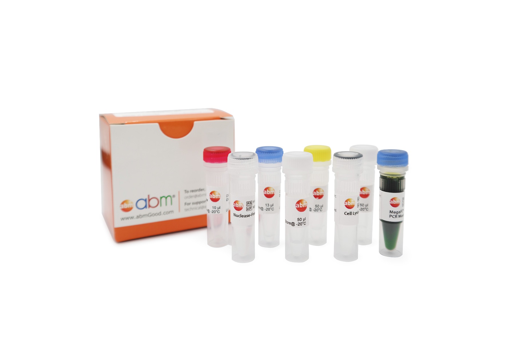
CRISPR Genomic Cleavage Detection Kit


| Cat. No. | G932 | ||||||||||||||
| Name | CRISPR Genomic Cleavage Detection Kit | ||||||||||||||
| Unit | 100 Reactions | ||||||||||||||
| Category | Molecular Biology Enzymes and Kits | ||||||||||||||
| Description |
abm’s CRISPR Genomic Cleavage Detection Kit is a simple and rapid assay designed to verify your genomic editing process. Precision is essential in genome editing and gene modification, and confirming successful gene editing early can save both time and resources. This kit allows you to easily detect genomic cleavage, ensuring accurate results in your experiments. The kit uses CRISPR-edited samples as templates in PCR reactions targeting your specific region of interest. It requires a pair of primers flanking each sgRNA target site to detect genomic cleavage, which can be ordered from abm (Cat. No. C336 - sgRNA PCR Primer Pair Design & Synthesis Service). After PCR amplification, the products are denatured and re-annealed, creating mismatches within the double-strand DNA. Our detection enzyme recognizes these mismatches and cleaves the strands to produce distinguishable band sizes upon gel analysis. The ready-to-use kit includes all necessary reagents, including a control template and primers to ensure reliable results. The control template produces bands of approximately 750bp after PCR amplification and 500bp and 250bp after enzymatic cleavage. With a quick processing time, this assay is a valuable addition to any genomic-editing workflow. Kit Features:
|
||||||||||||||
| Storage Condition |
Store all components at -20°C. |
||||||||||||||
| Material Citation | If use of this material results in a scientific publication, please cite the material in the following manner: Applied Biological Materials Inc, Cat. No. G932 |
| How does this kit verify gene editing? | |
|
The CRISPR Genomic Cleavage Detection Kit is designed to verify CRISPR-based genome editing by detecting mismatches in PCR products. After PCR amplification of CRISPR-edited samples, the products are denatured and re-annealed, creating mismatches at the edited site. The detection enzyme cleaves these mismatches, producing distinguishable fragments that can be analyzed on a gel. The kit ensures precision by confirming successful edits early in the process, saving time and resources.
|
| What should I expect to see on the gel when using the CRISPR Genomic Cleavage Detection Kit? | |
|
On the gel, you should expect the following:
|
| What primers are required for use with the CRISPR Genomic Cleavage Detection Kit? | |
|
The kit requires primers that flank the sgRNA target site for detecting genomic cleavage. These primers can be designed and ordered from abm’s sgRNA PCR Primer Pair Design & Synthesis Service (Cat. No. C336).
|
| What is the control template used for in this kit? | |
|
The control template serves as a reference to verify the performance of the assay. It produces PCR products of approximately 750 bp, and after cleavage, it produces bands of 500 bp and 250 bp. These control bands help confirm the assay’s accuracy.
|
| Can this kit be used for monoclonal CRISPR Knock-Out cell selection? | |
|
No, this kit is designed to detect the presence of genomic editing but is not suitable for monoclonal selection of CRISPR Knock-Out cells. For that purpose, you should use the Screen It™ CRISPR Cas9 Cleavage Detection Kit (Cat. No. G990).
|
| What should I do if I see non-specific bands on the gel? | |
|
If non-specific bands appear, try using the touchdown PCR method to improve specificity. Adjusting PCR conditions can reduce non-specific amplification, helping ensure clear and accurate results for cleavage analysis.
|
-
Chakraborty, A., Dutta, A., Dettori, L. G., Daoud, R., Li, J., Gonzalez, L., ... & Feng, W. (2024). Complex interplay between FMRP and DHX9 during DNA replication stress. Journal of Biological Chemistry, 300(1). https://doi.org/10.1016/j.jbc.2023.105572
Chakraborty, A., Dutta, A., Dettori, L. G., Li, J., Gonzalez, L., Xue, X., ... & Feng, W. (2021). FMRP directly interacts with R-loop and shows complex interplay with the DHX9 helicase. bioRxiv, 2021-04. https://doi.org/10.1101/2021.04.21.440759
Ortiz-Cuaran, S., Swalduz, A., Foy, J. P., Marteau, S., Morel, A. P., Fauvet, F., ... & Saintigny, P. (2022). Epithelial-to-mesenchymal transition promotes immune escape by inducing CD70 in non-small cell lung cancer. European Journal of Cancer, 169, 106-122. https://doi.org/10.1016/j.ejca.2022.03.038
Pizzoni, A., Zhang, X., Naim, N., & Altschuler, D. L. (2023). Soluble cyclase-mediated nuclear cAMP synthesis is sufficient for cell proliferation. Proceedings of the National Academy of Sciences, 120(4), e2208749120. https://doi.org/10.1073/pnas.2208749120
Saikia, B. B., Bhowmick, S., Malat, A., Rani, M. P., Thaha, A., & Abdul-Muneer, P. M. (2024). ICAM-1 deletion using CRISPR/Cas9 protects the brain from traumatic brain injury-induced inflammatory leukocyte adhesion and transmigration cascades by attenuating the Paxillin/FAK-Dependent Rho GTPase Pathway. Journal of Neuroscience, 44(11). https://doi.org/10.1523/JNEUROSCI.1742-23.2024
Santo-Domingo, J., Lassueur, S., Galindo, A. N., Alvarez-Illera, P., Romero-Sanz, S., Caldero-Escudero, E., ... & Wiederkehr, A. (2023). SLC25A46 promotes mitochondrial fission and mediates resistance to lipotoxic stress in INS-1E insulin-secreting cells. Journal of Cell Science, 136(8), jcs260049. https://doi.org/10.1242/jcs.260049
Smeir, M., Chumala, P., Katselis, G. S., & Liu, L. (2024). Lymphocyte-Specific protein 1 regulates expression and stability of endothelial nitric oxide synthase. Biomolecules, 14(1), 111. https://doi.org/10.3390/biom14010111
Zhang, X., Tanwar, V. S., Jose, C. C., Lee, H. W., & Cuddapah, S. (2022). Transcriptional repression of E‐cadherin in nickel‐exposed lung epithelial cells mediated by loss of Sp1 binding at the promoter. Molecular carcinogenesis, 61(1), 99-110. https://doi.org/10.1002/mc.23364